News
3D printing bone tissue
Jun 28 2022
Read More
Ultrasound imaging research continues to push forward, aiming to create new frameworks for medical impact with recent findings captured in a new publication in Ultrasound in Medicine & Biology.
Dr Andres Ruland
The paper, led by ACES and University of Wollongong (UOW)’s Dr Andres Ruland, highlights the validation of signal processing methods for ultrasonic imaging, with a focus on biological constructs. The research was assisted by ACES Director Prof Gordon Wallace as well as Prof Jim Hill from the University of South Australia, who supervised the computation involved in the work.
Dr Ruland has been focusing on the area of ultrasound for several years and presented on its technology as a non-destructive technique for biomaterial evaluation during last year’s ACES Technology Showcase (watch below).
To find out more about ultrasound imaging, the research captured in this new publication and its future implications, we spoke with Dr Ruland.
Can you explain this new research in more detail?
Our work looked at validating our signal processing method for the ultrasonic imaging of biological constructs with dynamic properties, referring to a construct with varying morphology and composition.
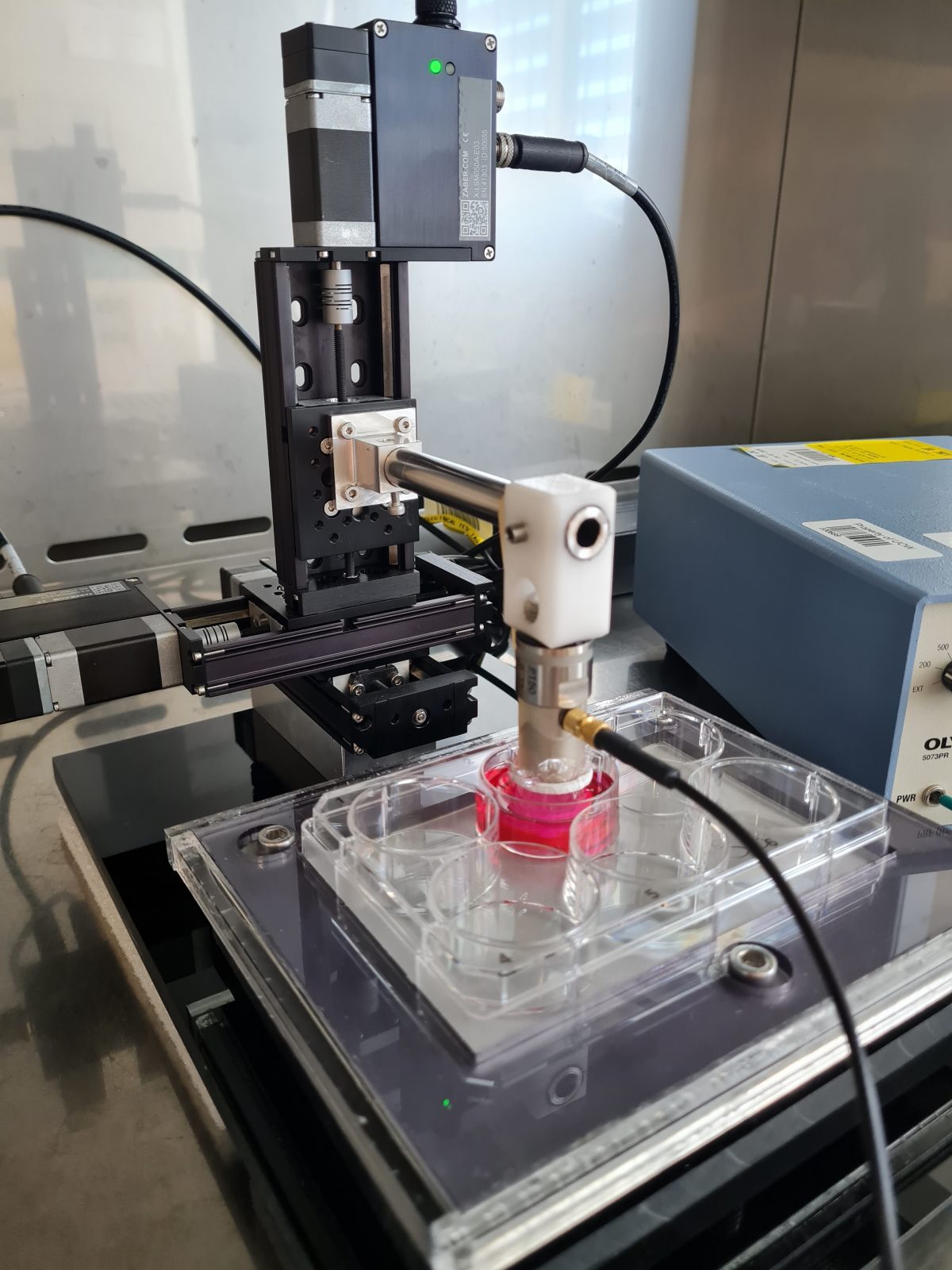
In biological constructs, such physical changes originate because of the metabolic activity of stem cells laden into a biodegradable construct. These types of constructs are being used in biological research for recreating physiological processes and structures on the bench (e.g. in-vitro). The advantage of ultrasound is that it allows the mapping and quantification of biological constructs in detail and non-invasively.
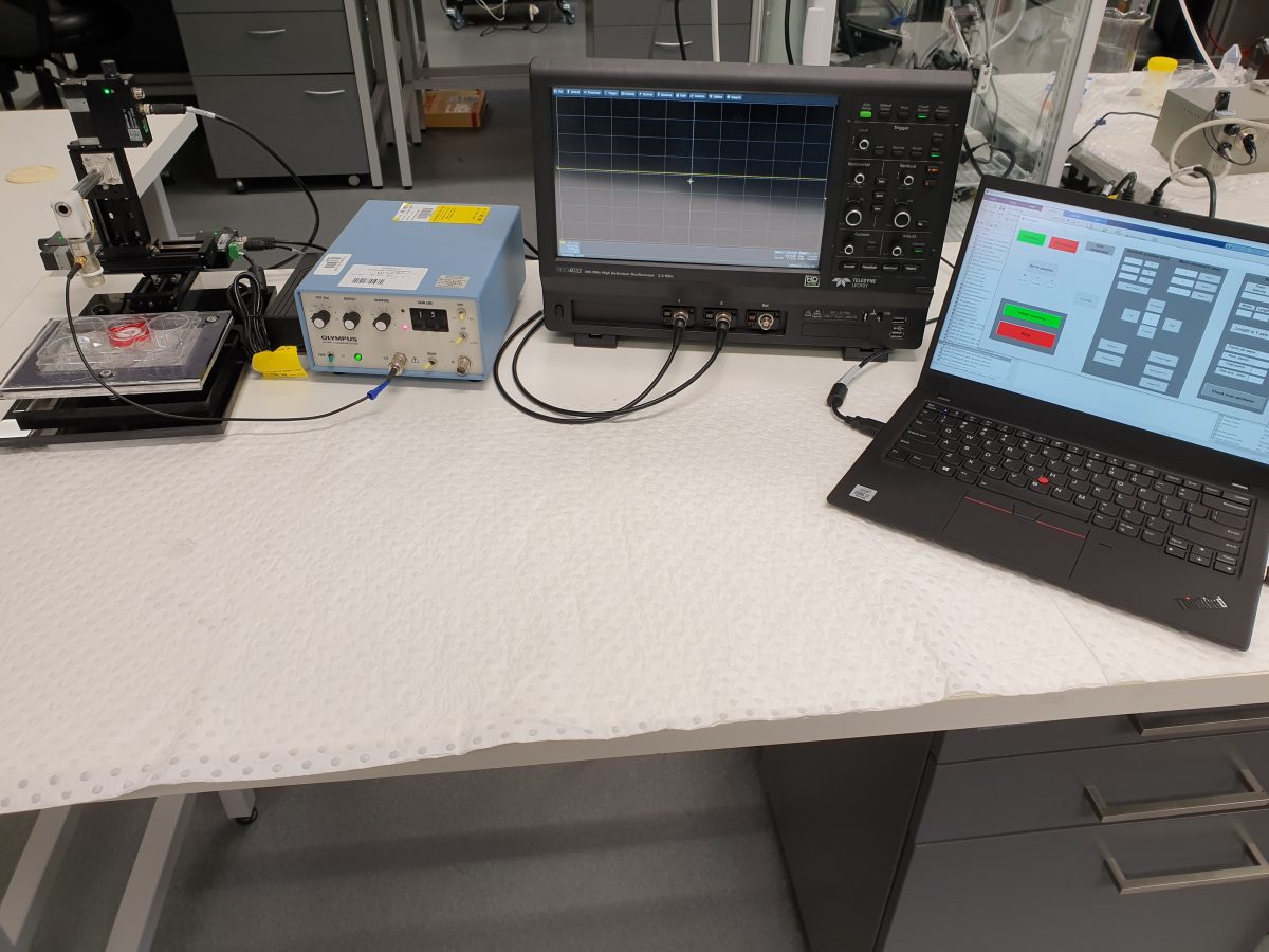
Another aspect to point out is that we specialised on the use of a reference phantom method for the processing of ultrasound images, where the unknown sample is calculated with respect to a phantom of known properties. The challenge is to explore whether the physical changes occurring in biological constructs with dynamic properties may alter the ultrasonic determination of the constructs, and if so, how can it be compensated. This was studied by designing a set of strategic experiments using acellular constructs for mimicking the conditions that may be found in biological constructs of dynamic properties, where the morphology sound speed and attenuation are expected to change significantly.
We found that sound speed increase had a significant effect on the ultrasound intensity, and we developed an empirical function for the compensation of such effect. Fortunately, we found that our method was robust enough for compensating morphology changes as well as large attenuation changes with respect to the reference phantom.
Overall, with the introduction of the sound speed compensation function, we are now able to evaluate any biological construct with respect to a phantom independently of its sound speed. This is a great advantage and a step towards the standardisation of ultrasound, where instead of preparing a different phantom depending on the sample under evaluation, we can have a generic phantom. This is advantageous not only in practical terms, but also important since the ultrasound estimates are dependent on the phantom used.
You worked with Prof Jim Hill from the University of South Australia for this research. How did the collaboration come about?
This work was challenging because of all the equations involved. We were very fortunate to have Prof Jim Hill from the University of South Australia as a collaborator for supervising the computation involved. We are hoping that we will be able to conduct further collaborations with him. It is important to establish a network of researchers with diverse skills in order to expand and strengthen our capabilities.
What are the future implications for this research and ultra-imaging?
The work we have published provides the theoretical framework and validation for conducting other studies of medical impact.
Another interesting project we are working on with the University of Melbourne is the monitoring of cartilage growth in a construct using ultrasound. The advantage of ultrasound is the possibility to non-invasively map and quantify the cartilage development within the full construct’s dimension and depth. This is an important aspect to highlight since optic-based techniques, which are the gold standard for biological construct evaluation, are only able to map shallow regions of the construct while destructive procedures such histology is needed for viewing the construct in its full dimension.
The cartilage project has now concluded and has been submitted to a peer-reviewed journal of high impact factor. We are very excited about such work as it will bring the use of ultrasound for tissue engineering to a new standard. Other applications being currently conducted is the use of ultrasound for monitoring wound healing, and is providing very encouraging results. Hopefully, we will be able to provide an update in the following months.
Thanks, Andres and well done. Access the paper here.